
An international research collaboration led by physicists from the University of Southampton has demonstrated a non-invasive optical method to access quantum physics in artificial lattices.
The new study builds on increasing interest over the past decade in optically responsive quasiparticles known as polaritons that originate from the strong-coupling regime between light and matter.
The results could inspire the creation of field-programmable polariton circuitry and new strategies for the robust confinement of coherent light sources, such as lasers.
Researchers from Southampton's Hybrid Photonics group, Lancaster University and the Skolkovo Institute of Science and Technology (Skoltech) in Russia have published their findings in the journal Nature Communications.
Creating artificial lattices for quantum particles enables scientists to explore physics in an environment which might not be conventionally found in nature. Artificial lattices are especially appealing since their symmetries can lead to exactly solvable models and transparent understanding of their properties.
Designing them is, however, a challenging task with limited flexibility as most methods require materials to be irreversibly engineered to get the job done.
The new research, led Southampton’s Dr Lucy Pickup, Dr Helgi Sigurdsson and Professor Pavlos Lagoudakis, and Lancaster's Professor Janne Ruostekoski, overcame this challenge by using structured laser light to synthesise arbitrarily shaped and reprogrammable artificial lattices. These cavity-polariton systems could be changed from one lattice to another without the costly need to engineer a new system from scratch.
Dr Pickup, article co-author, says: "The results open a path to study dissipative many-body quantum physics in a lattice environment with properties that cannot be reproduced in normal Hermitian quantum systems."
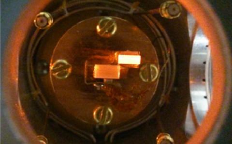
When laser light hits a semiconductor quantum well it excites a lot of electrons, holes and bound states of the two, known as excitons. When the quantum well is put between two mirrors, forming a trap (or a cavity) for the photons, some of the exciton particles start becoming 'dressed' in photons, forming new part-light part-matter quasiparticles known as exciton-polaritons or cavity-polaritons.
Exciton-polaritons are very interactive and bounce frequently off one another. However, they also bounce off normal electrons, holes, and excitons in the background around them.
By applying laser light in a geometrically structured fashion the exciton-polaritons start bouncing off the excited electrons, holes and excitons following the shape of the laser. In other words, the exciton-polaritons experience a synthetic potential landscape imprinted by the laser.
The laser generated potential landscapes are only felt by the exciton-polaritons and not the photons inside the cavity, making the system uniquely different from photonic crystals. By creating a laser pattern with translational symmetry the researchers produced the fundamental signature of solid state systems, the formation of crystal energy bands for exciton-polaritons, just like one would observe for electrons in solid state materials.
The produced bands can be reconfigured by simply adjusting the laser pattern, making the method applicable to a variety of potential applications from optical-based communications and information processing, to high sensitivity detectors in biomedicine and topologically protected lasers. The results also open a path to study fundamental many-body lattice physics in an open quantum environment.
Dr Sigurdsson, article co-author, adds: "This is an exciting development for the relatively new field of non-Hermitian topological physics and has only just begun."
Articles that may also interest you
Researchers at the University of Southampton have visualised a healthy mouse kidney with high spatial resolution and high contrast using a promising, emerging X-ray imaging technique.
The new study, which was led by scientists in Physics and Astronomy, highlights X-ray speckle-based imaging's (SBI) potential as an easily implemented and non-invasive approach for next generation biomedical scanning using inexpensive optical elements.
SBI imaging is capable of delivering qualitative information, such as tissue micro-structure, and quantitative information, such as tissue density, by merely adding a piece of sandpaper to a conventional X-ray imaging setup.
Scientists have published their findings this month in The Optical Society's flagship journal Optica.
Studying the microscopic structure of tissues is essential for a better understanding of organ functions and can, moreover, be key to identifying the cause, stage and progress of treatment of an illness. Conventional histology, the current gold standard for soft-tissue visualisation, requires several days of preparation and destroys samples so they cannot be used for further analysis. The process can also only produce 2D images that make it hard to accurately measure or analyse sample structures and their connectivity in 3D.
X-ray imaging can penetrate deeply into samples and visualise their inner structure without having to physically slice them, while also reconstructing a 3D volume by taking images at different rotation angles.
In X-ray phase-contrast imaging, scientists measure the refraction of the X-rays when passing through the sample. Compared to conventional X-ray imaging based on X-ray absorption, this approach is much more sensitive to small changes in density that are present in biological soft tissue.
This new research, led by Dr Marie-Christine Zdora, Dr Irene Zanette, and Professor Pierre Thibault, now of the University of Trieste, advances the exploration of SBI imaging beyond initial proof-of-principle demonstrations to an important real-world application.
The technology’s potential for virtual histology, where a sample is virtually sliced on a computer along a desired plane, is exhibited using a whole unstained, hydrated kidney of a healthy mouse imaged at a synchrotron, a large-scale facility that produces extremely bright X-rays.
Dr Zdora, Research Fellow and lead author of the study, says: "It seems hard to believe that simply adding a piece of sandpaper to a standard X-ray imaging setup can lead to such beautiful results and our medical collaborators were amazed by the image quality of our technique.
"Achieving such detail and contrast in hydrated biological soft tissue without the use of any staining agents is extremely challenging. The high image quality combined with the simple, cost-effective experimental setup make our method very promising for future applications in histopathology and biomedical research, but also in other fields such as materials science.
"At Southampton, we are currently working on implementing our technique at a laboratory X-ray source, which will make it more accessible to a wider range of users."
The latest project builds upon six years of SBI research by the authors, four of them at Southampton, and was carried out under Dr Zanette's Royal Society University Research Fellowship and Enhancement Award.
X-ray imaging took place at the ESRF (European Synchrotron Radiation Facility) in France, with conventional histology carried out on the same sample for comparison at the University Hospital Southampton’s Biomedical Imaging Unit. Samples were prepared at the University of Zurich.
The project's funding sources included the Royal Society, the European Research Council and the Swiss National Science Foundation.
The team are next planning pre-clinical studies on medical conditions that will image healthy and diseased tissue before comparing the results. SBI will allow for measuring a large number of samples at high throughput with minimal sample preparation. The extracted 3D data volumes can then be analysed with automated algorithms to extract the desired information on the tissue. This will enable large-scale studies on a wide range of diseases in a much shorter timeframe and with less manual input than conventional histology.
The scientists are also extending the method to other fields such as materials science, and palaeontology.
Articles that may also interest you
Research
Research interests
optical levitation system, optical sensor, metasurface
Publications
Sun, Chuang and Yan, Jize (2022) A hybrid method to calculate optical torque: application to a nano-dumbbell trapped by a metalens. AIP Advances, 12 (7), [075024]. (doi:10.1063/5.0094665).
Contact
Telephone: +44 (0) 23 8059 7864761523

Quantum Technologies for Fundamental Physics
Our research addresses fundamental questions in the overlap of quantum theory and general relativity using quantum information and metrology techniques. We study how to use quantum systems to measure gravitational waves and set constraints on dark energy
Hybrid Quantum Networks Lab
Our research interests lie in quantum light-matter interactions between atomic ensembles and single photons, to form large-scale quantum photonic networks for the processing of quantum information over global distances.
An international study led by the University of Southampton has demonstrated for the first time how light can be used to glue together negative charges and create a novel form of matter.
The ground-breaking research, led by Physics and Astronomy's Professor Simone De Liberato, opens new possibilities for engineering novel artificial atoms with designer electronic configurations.
Researchers built upon a theoretical prediction from 2019 to fabricate a nanodevice that trapped electrons within nanoscopic wells.
When photons struck the device with a high enough energy they would extract electrons from the wells. The team then enclosed the device between two gold mirrors, dramatically increasing the interaction between light and matter.
The study observed that a negatively-charged electron would remain trapped, bound to the other negatively-charged electrons in a novel electronic configuration like a 'subatomic zip tie' stabilised by the photon.
Scientists have published their findings in the journal Nature Physics.
The technique will be used to broaden the catalogue of materials available to design photonic devices.
Read the full story on the main news page.
Articles that may also interest you
Publications
Vinante, Andrea, Gasbarri, Giulio, Timberlake, Christopher, Toros, Marko and Ulbricht, Hendrik (2020) Testing dissipative collapse models with a levitated micromagnet. Physical Review Research. (In Press)
Fadeev, Pavel, Timberlake, Chris, Wang, Tao, Vinante, Andrea, Band, Yehuda B, Budker, Dmitry, Sushkov, Alexander O, Ulbricht, Hendrik and Kimball, Derek F Jackson (2021) Ferromagnetic gyroscopes for tests of fundamental physics. Quantum Science and Technology, 6 (2), [024006]. (doi:10.1088/2058-9565/abd892).
Timberlake, Christopher, Vinante, Andrea, Shankar, F, Lapi, Andrea and Ulbricht, Hendrik (2021) Probing modified gravity with magnetically levitated resonators. Physical Review D, 104 (10), [L101101]. (doi:10.1103/PhysRevD.104.L101101).
Researchers from the University of Southampton have demonstrated a new material family that will revolutionise optical circuits to replace parts of traditional electronic hardware.
The materials allow rapid reversible switching between two states, known as phase change, which has previously been limited to electronic circuits as standard commercially available materials suffer from large optical losses.
Scientists from the Quantum, Light and Matter group and Optoelectronics Research Centre (ORC) have designed the phase change materials to exhibit no loss of light at telecommunication wavelengths and be switched with very low power.
The technology is compatible with existing silicon photonic circuits and opens the door for more advanced applications. Researchers have published their findings in Advanced Functional Materials.
Lead authors Dr Matthew Delaney and Dr Ioannis Zeimpekis pinpointed the material structure and composition to enable high transparency while exhibiting low power modulation of light. They found that the new composition has 100 times less loss than the current state-of-the-art optical materials.
Their material was deposited on top of optical chips, where a short laser pulse was used to crystalize the material and change the phase of the guided light. The researchers demonstrated this property reversibly thousands of times. Importantly, the material remembers its last state without any applied signals, leading to large potential power savings.
Professor Otto Muskens, Head of the Integrated Nanophotonics group, says: “This new technology will simplify and enable newly emerging applications such as solid-state LiDAR, quantum and neuromorphic computing that are currently limited by the performance of the existing materials.
“Neuromorphic and programmable photonics are set to revolutionise the industry as they offer new paradigms for data processing going far beyond existing hardware. Quantum optical circuits are on the horizon and ultralow loss components are needed to make the next step in controlling and routing quantum information.”
Traditional communication electronics consume a huge proportion of their energy at the interconnection level, and their bandwidth is directly limited by the communication length. Using photons instead of electrons alleviates these shortcomings.
Phase change photonics offer much promise for the future of integrated silicon photonic circuits, with some of the world’s largest companies competing in the race for fully integrated optical solutions. However, the high absorption losses in current commercially available materials have prevented their use in larger photonic systems such as in interconnects between data servers, where the photonic technology is projected to excel.
Professor Dan Hewak, ORC co-author who has spent decades on phase change materials, says: “This is a significant breakthrough for optoelectronics. Our team has now demonstrated a material which bridges the gap between electronics and photonics and we expect to see further advances resulting from their discovery.”
The new phase change family has been designed as part of a series of research projects funded by the Engineering and Physical Sciences Research Council (EPSRC), including ChAMP, the Manufacturing and Application of Next Generation Chalcogenides (EP/M015130/1), Cornerstone (EP/L021129/1), The Physics and Technology of Photonic Metadevices and Metasystems (EP/M009122/1) and Nanostructured photonic metamaterials (EP/G060363/1). The new materials reside within the chalcogenide family as they combine antimony and sulfur or selenium.
The team is currently working to implement more photonic circuit components with the aim to design a neuromorphic computing photonic chip with in-memory computing capabilities. It is expected that this method will replace current technologies within the next couple of years enabling a leap forward for the technology of photonic computing.